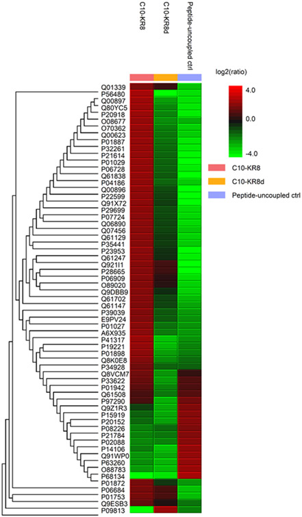
Figure 1.
Mass spectrometry analysis of mouse serum proteins binding to the L- and D-forms of C10-KR8 immobilized to beads (see Methods). Heat map representation of the abundances of 20 significantly up-regulated (red) and down-regulated (green) proteins (PEAKS Q) bound to C10-KR8, C10-KR8d, and the peptide uncoupled control samples (left to right) after unsupervised hierarchical clustering.

