
Figure 1. Alveolar simplification and loss of gas exchange surface after perinatal inflammatory injury.
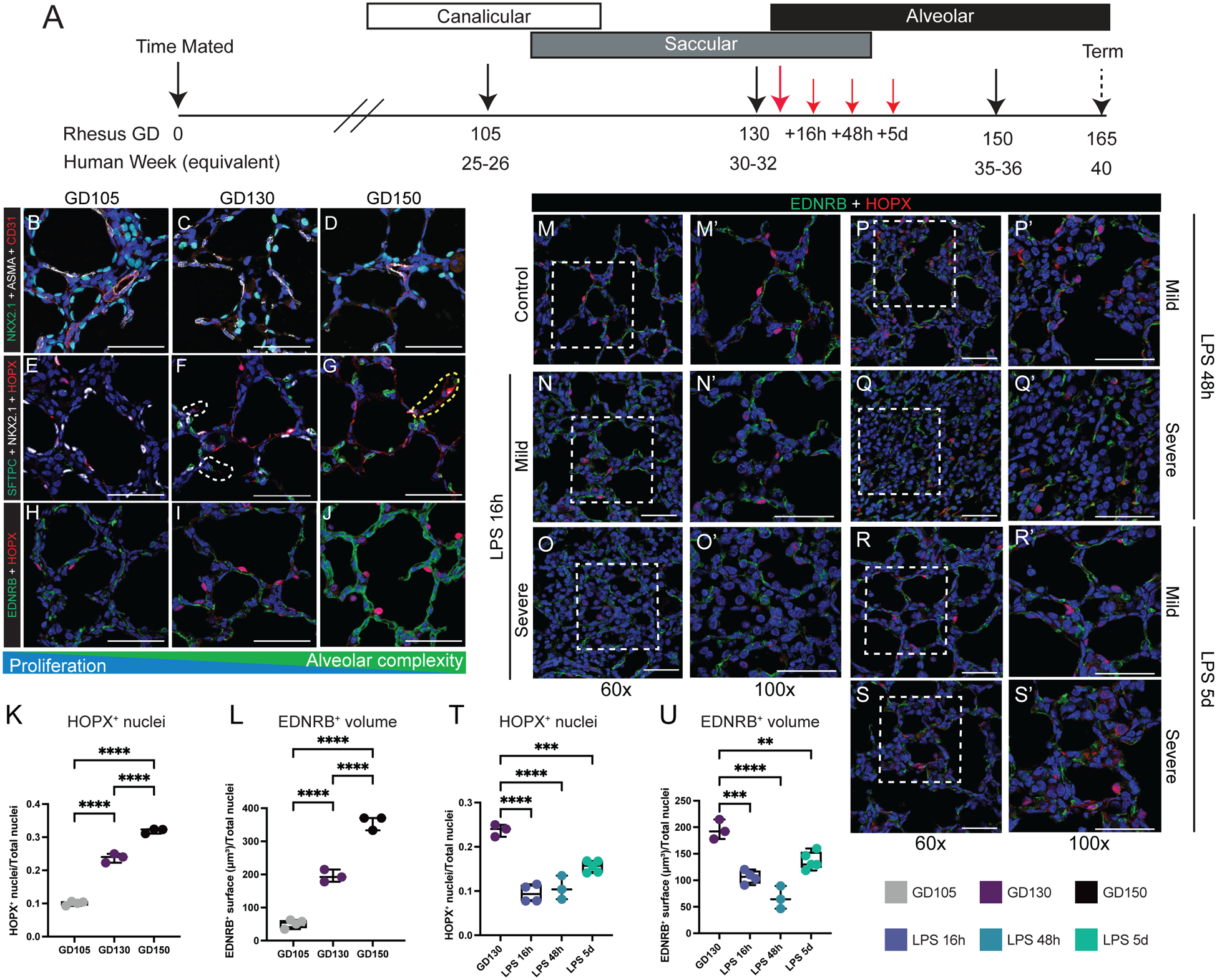
(A) Experimental design. Control animals obtained at at GD105 [canalicular; n=3], GD130 [saccular; n=11], and GD150 [alveolar; n=3] (black arrows). For injury, rhesus macaque dams received intra-amniotic LPS injection at GD130, with subsequent fetal delivery by C-section at 16h (n=8), 48h (n=3), or 5d (n=7) after injury (red arrows). (B-D) NKX2.1+ epithelial cells (green) form close interactions with both ASMA+ myofibroblasts (white) and CD31+ endothelial cells (red). (E-G) Epithelial differentiation of HOPX+ AT1 cells (red) and SFTPC+ AT2 cells (green) increase as development progresses. (H-J) Interactions of HOPX+ AT1 cells (red) and EDNRB+ alveolar capillary endothelial cells (ACs; green) increase during patterning of the developing gas exchange surface. K-L) Quantification of HOPX+ nuclei (L) and EDNRB+ surfaces (L) at each stage of lung development. (M-S) Extensive disruption of gas exchange surface, shown through the disordered patterning and interactions of HOPX+ AT1 cells (red) and EDNRB+ ACs (green) in both mild and severely injured regions up to 5d following LPS. T-U) Quantification of HOPX+ nuclei (T) and EDNRB+ surfaces (U) during injury. Scale bars = 50 μm. Full list of samples and treatment groups in Table S1. ** = p <0.01, *** = p <0.001, **** = p < 0.0001 by Kruskal-Wallace test.