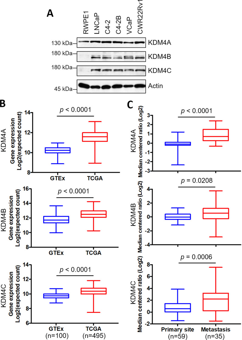
Fig. 1.
KDM4A, KDM4B, and KDM4C are overexpressed in PCa. A Expression of KDM4A, KDM4B, and KDM4C in an immortalized prostatic epithelial cell line (RWPE1) and PCa cell lines (LNCaP, C4-2, C4-2B, VCaP, and CWR22Rv1) using Western blotting analysis. Actin served as an internal control. B Expression of KDM4A, KDM4B, and KDM4C in PCa (TCGA) and normal (GTEx) specimens based on the UCSC Xena tool (https://xenabrowser.net). C Analysis of expression of KDM4A, KDM4B, and KDM4C in primary and metastatic PCa tissues based on the Grasso prostate dataset in Oncomine database (https://www.oncomine.org/resource/login.html). Statistical significance was determined using Student's t-test

