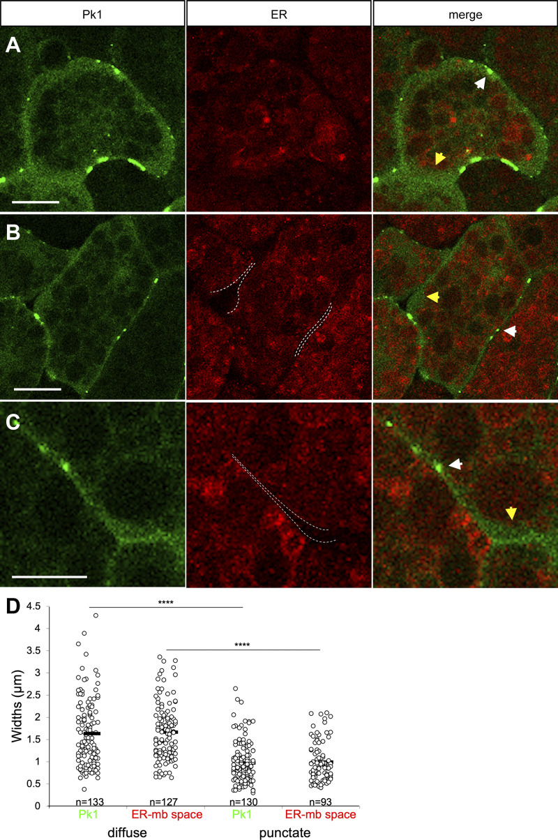
Figure S3.
Pk1 is enriched at the ER-free cell periphery in PCM tissue mosaically labeled with Pk1-venus and ER-Tracker Red. (A–C) PCM explants labeled with Pk1-venus and ER-Tracker Red. Dashed lines indicate ER domain boundaries. White arrows, Pk1 puncta and plaques; yellow arrows, puncta-less regions of cell periphery. Scale bars, 10 μm. (D) Widths of Pk1 zone and ER-membrane space in PCM explants. At diffuse Pk1: Pk1, n = 133; ER-membrane space, n = 127. At puncta: Pk1, n = 130; ER-membrane space, n = 93. n, number of cells. ****, P ≤ 0.0001 in a two-tailed Student’s t test. Data distribution was assumed to be normal but was not formally tested.

