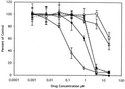
FIG. 6.
Inhibition of the development and multiplication of intracellular C. parvum parasites by lipophilic antifolates. MDCK epithelial cells were plated, grown, and infected as described in the legend to Fig. 5. Following postinfection washes to remove unexcysted oocysts and extracellular sporozoites, deficient medium–1% dFCS was added to each well of the chamber slide and 20 mM stock solutions of the indicated antifolate drugs were serially diluted fivefold across seven wells. The last well received no drug and served as a control to quantify uninhibited growth. Drugs and media were renewed at 24 h postinfection, the experiment was terminated at 48 h, and the monolayers were processed as described in the legend to Fig. 5. The slides were microscopically examined for epifluorescence, and the numbers of parasite-containing fluorescent vacuoles were counted in each of five microscopic fields per well (magnification, ca. ×400). Results are expressed as percentages of the control determined as follows: (average number of parasite-containing vacuoles in drug-treated cells ÷ average number of vacuoles in drug-free controls) × 100. Error bars indicate the standard deviation at each drug concentration. Each drug was tested at least twice with similar results. Control experiments using uninfected MDCK cells showed that the drug concentrations used were not toxic to the cells (data not shown). Symbols: ◊, trimetrexate; □, trimethoprim; ▵, pyrimethamine; ×, compound V.10; ∗, compound V.15; ●, compound V.16.

