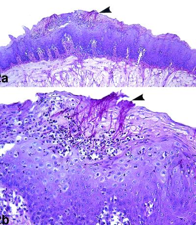
FIG. 2.
Oral candidiasis in control immunosuppressed rats. PAS staining was used. (a) The lesional mucosa showed extensive hyperplasia of the epithelium of the dorsum of the tongue, with numerous C. albicans organisms (arrowhead) in the epithelium. Magnification, ×100. (b) Detail of panel a, showing necrosis of the superficial epithelium with intraepithelial microabscesses and numerous candidal hyphae (arrowhead). Magnification, ×400.

