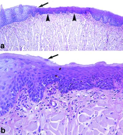
FIG. 3.
Oral candidiasis in immunosuppressed rats treated with GW471558 at 10 mg/kg. PAS staining was used. (a) Extensive lingual epithelium atrophy (arrowheads) compared to the normal lingual epithelium of the tongue (arrow) is shown. Magnification, ×100. (b) The atrophic epithelium shows an increase of basal and suprabasal keratinocyte proliferation with incomplete differentiation of the scamous and corneum stratum. The limit between normal and lesional epithelium is very pronounced (arrow). C. albicans was not observed in all lingual mucosa. Magnification, ×400.

