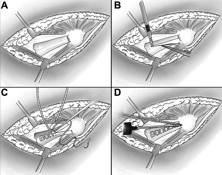
Figure 1.
(A) Wide exposure via the muscle-splitting approach showing the proximal avulsion of the ulnar collateral ligament (UCL) in a right elbow. (B) The graft was split longitudinally but sparing the origin of the intact distal attachment. The UCL graft was examined for any areas of attenuation of calcifications that would preclude a repair. (C) The UCL graft was sutured in a running fashion starting at the nonruptured end, tensioning the suture after each throw. The final pass of each suture limb ended with the tails exiting superficially to the graft. (D) Both free ends of the suture were secured to the anatomic bony footprint with a knotless suture anchor.

