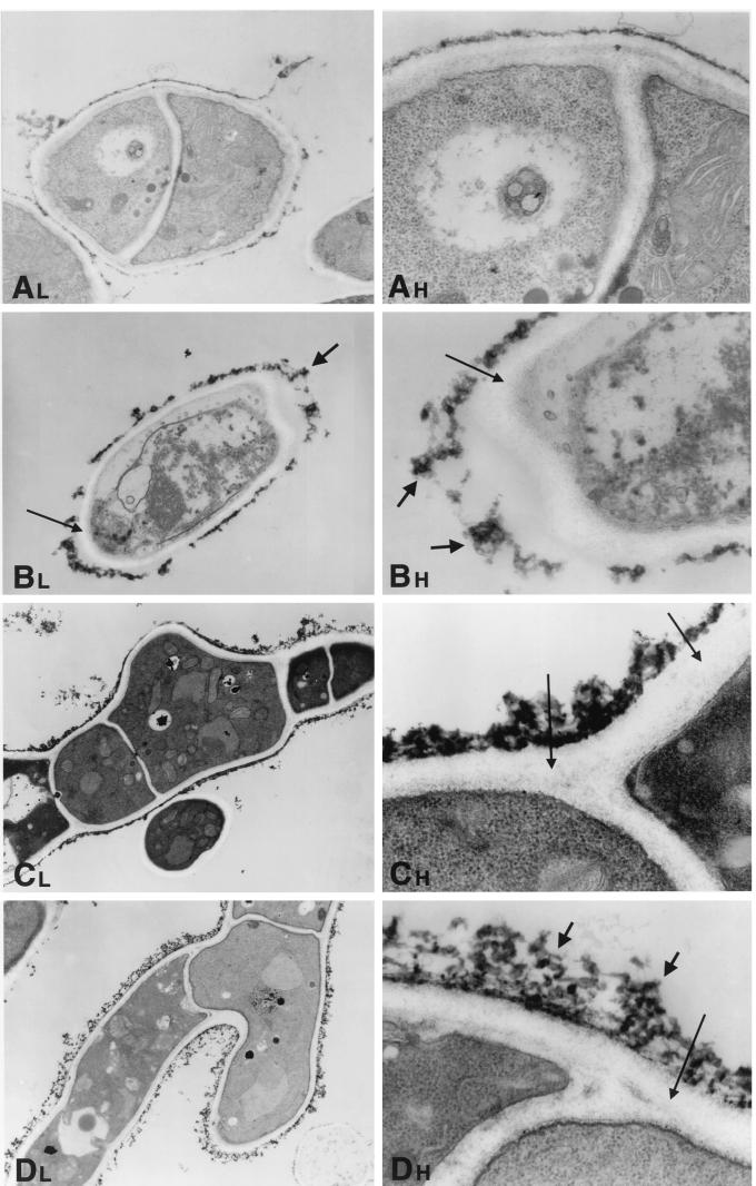
FIG. 6.
Time-sequenced electron microscopic changes of A. fumigatus (isolate 4215) after 8 h of incubation with NZ (32 μg/ml) and FK (0.5 μg/ml) alone and in combination. L, lower magnifications; H, higher magnifications. (A) Normal growth control of A. fumigatus hyphae. Magnifications, ×1,100 and ×3,100. (B) NZ alone. The cell membrane has undergone significant changes, with loss of the electron-dense lipid bilayer structure. Small microvesicles may be seen in the area of the cell membrane. The inner fibrillar layer of the cell wall has lost its characteristic distinct, fine granularity. This layer is also much wider than that of controls (thin arrows). The outer fibrillar layer of the cell wall is aggregated and thicker than in the controls (thick arrows). In other areas, the outer fibrillar layer appears to be detached from the cell itself. Magnifications, ×900 and ×3,100. (C) FK alone. The outer fibrillar layer is focally thicker than that of the controls, and there are aggregates similar to those seen in the NZ-treated cells. The granularity of the inner fibrillar layer is relatively preserved, but it is not as fine as in the controls (arrows). Magnifications, ×620 and ×4,800. (D) NZ plus FK. The outer fibrillar layer has a lattice-like structure that is loose and thready (thick arrows). The inner fibrillar layer is not uniformly visible at these magnifications (thin arrow). Magnifications, ×620 and ×4,800.

