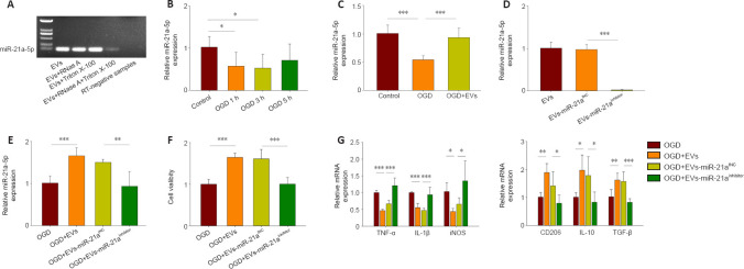
Figure 4.
miR-21a-5p in MSCs-EVs attenuates the inflammatory response after OGD in BV-2 cells.
(A) RT-PCR analysis of the levels of miR-21a-5p in EVs, EVs pretreated with RNase A, EVs pretreated with Triton X-100, EVs pretreated with Triton X-100 and RNase A, and RT-negative samples. (B) qRT-PCR analysis of the levels of miR-21a-5p in BV-2 cells after 1-, 3-, and 5-hour OGD followed by 24-hour reoxygenation. The control group was cultured normally without OGD. (C) qRT-PCR analysis of the levels of miR-21a-5p in the presence or absence of MSCs-EVs. The cells had undergone 3-hour OGD followed by 24-hour reoxygenation. (D) qRT-PCR analysis of the levels of miR-21a-5p in the different EVs (EVs, EVs-miR-21aINC, and EVs-miR-21ainhibitor). (E) qRT-PCR analysis of the levels of miR-21a-5p following treatment with EVs, EVs-miR-21ainhibitor, or EVs-miR-21aINC. The cells had undergone 3-hour OGD followed by 24-hour reoxygenation. (F) The viability of BV-2 cells, assessed using CCK8, following treatment with EVs, EVs-miR-21ainhibitor, or EVs-miR-21aINC. The cells had undergone 3-hour OGD followed by 24 -hour reoxygenation. (G) qRT-PCR analysis of the mRNA levels of TNF-α, IL-1β, iNOS, CD206, IL-10, and TGF-β for cells treated with no EVs, EVs, EVs-miR-21aINC, or EVs-miR-21ainhibitor. The cells had undergone 3-hour OGD followed by 24-hour reoxygenation. Graphs show the mean ± SD. The experiments were carried out on six separate samples. *P < 0.05 (independent samples t-test) in B; *P < 0.05, ** P < 0.01, *** P < 0.001 (one-way analysis of variance with Bonferroni correction) in C–G. EV: Extracellular vesicle; IL: interleukin; iNOS: inducible nitric oxide synthase; MSC: mesenchymal stromal cell; OGD oxygen-glucose deprivation; qRT-PCR: quantitative reverse transcription-polymerase chain reaction; RT-PCR: reverse transcription PCR; TGF-β: transforming growth factor-β; TNF-α: tumor necrosis factor α.

