
Figure 3.
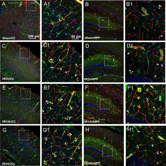
Changes in the expression of the pericyte marker PDGFR-β at different time points after cerebral ischemia/reperfusion.
(A–G) Representative photographs of the cerebral cortex (CC; A, C, E, G) and hippocampus (HIPP; B, D, F, H) showing platelet-derived growth factor receptor beta (PDGFR-β; green, Alexa Fluor 488)-labelled pericytes and CD31 (red, Alexa Fluor 594)-labelled blood vessels in the control (A and B), and 2 (C and D), 12 (E, F), and 24 hours after ischemia/reperfusion (G, H). (A1–H1) Magnified photographs of the boxed areas in panel A–H showing PDGFR-β and CD31 labeling in detail. In the sham group, pericytes had a conspicuous protruding ovoid cell body with long thin processes that coursed along the capillary for long distances and were embedded within the basement membrane. After 2, 12, and 24 hours of cerebral ischemia/reperfusion, the morphology of the capillaries near the pericyte cell bodies (shown by white arrows) changed, with distortion and even narrowing of the tube diameter (shown by yellow arrows), which positively correlated with the reperfusion time. The constriction of pericytes on capillaries in all model mice presented a similar pattern. The experiments were repeated six times. Scale bars: 200 μm in A–H, and 50 μm in A1–H1. CC: Cerebral cortex; DAPI: 4′,6-diamidino-2-phenylindole; PDGFR-β: platelet-derived growth factor receptor beta; HIPP: hippocampus.