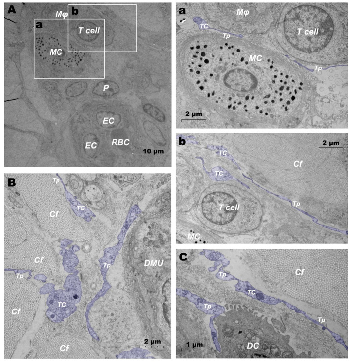
Figure 7.
Ultrastructural analysis of TCs in the steady state via TEM. (A) Location of TC in the DMUs in the steady state; (a) is the enlargement of the mast cell and surrounding TCs in (A); (b) is the enlargement of T cells and surrounding TCs in (A); (B) TCs surrounds the DMUs; (C) TC between collagen fibers and dendritic cells. RBC, red blood cell; EC, endothelial cell; P, Pericyte; MC, mast cell; Mϕ, macrophage; TC, telocyte; Tp, telopode; Cf, collagen fiber; DMU, dermal microvascular unit; DC, dendritic cell; Blue mark, telocytes. The scale bar (A) = 10 μm; (B) (a,b) = 2 μm; (C) = 1 μm.

