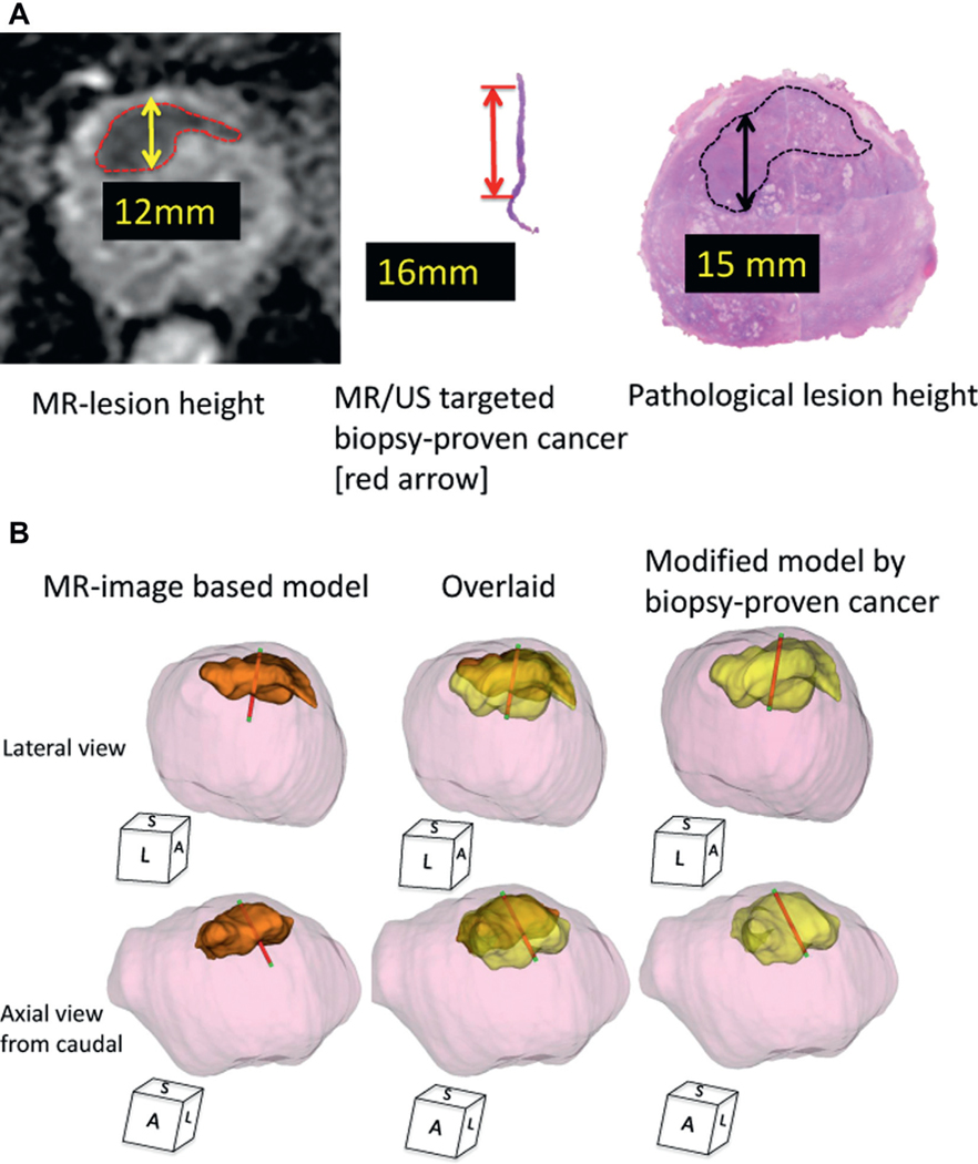
Figure 2.
Case presentation of MR visible lesion modification using MR/US fusion biopsy proven cancer core length. A, MR, targeted biopsy and prostatectomy data on 69-year-old man with PSA 7.4 ng/ml in whom prebiopsy MRI demonstrated PI-RAD level 5 suspicious lesion in anterior prostate. Height was 12 mm on DWI-ADC and MR/US image fusion targeted biopsy showed Gleason 3 + 4 with 16 mm core length. Subsequent robot-assisted RP revealed pathological T2c prostate cancer with dominant anterior cancer with maximum 15 mm anteroposterior lesion height. B, 3D tumor model, MRI based model and modified model created by replacing MR estimated lesion height with MR/US targeted biopsy proven cancer core length and vertically stretching height of original MRI based 3D model according to cancer core length. Final PCV of this index lesion was 4.5 ml. Model of 3D MCV (orange area) was 2.5 ml (56%). Newly developed, modified MCV (yellow area) was 3.6 ml (80%). Overlaid, MR/US targeted biopsy trajectory overlay of both 3D models using documented digitalized coordinates obtained from real-time 3D TRUS tracking technology with Urostation. Red areas indicate pathologically proven cancer tissue. Green areas indicate benign tissue. S, sagittal. L, lateral. A, anterior.

