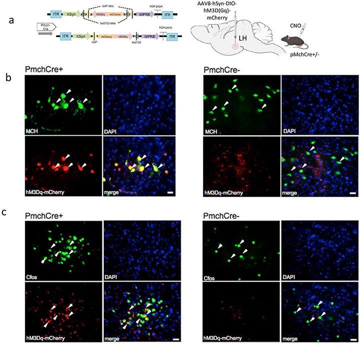
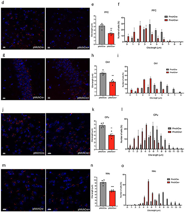
Fig. 4.
Chemogenic excitation of MCH shortens cilia length. a Experimental approach. Adult PmchCre + and PmchCre− mice underwent bilateral stereotaxic injection in the lateral hypothalamus with AAV-hSyn-DIO-hM3D(Gq)-mCherry to express DREADD in Cre-expressing MCH neurons. Clozapine-N-oxide (CNO) was delivered intraperitoneally to stimulate MCH neurons. Diagram was created with BioRender.com webpage. b Co-expression of mCherry fluorescence (red) identifying DREADD-expressing neurons and MCH immunofluorescence (green) and counterstained with DAPI (blue) in Pmch-Cre+and PmchCre− mice (scale bar = 10 μm, magnification = 40 ×). c mCherry fluorescence (red) identifies DREADD-expressing neurons, and c-Fos immunofluorescence (green) identifies cells recently activated in PmchCre + and PmchCre− mice (Scale bar = 10 μm). d, g, j, m Immunostaining of ADCY3 labeled in red fluorescence in the d PFC, g CA1, j CPu and m NAc via CNO-dependent activation of Gq signaling in PmchCre + and PmchCre− mice (scale bar = 10 μM). e, h, k, n) Quantification of the length of ADCY3 + primary cilia in the e PFC, h CA1, k CPu, and n NAc, **P < 0.01 and ***P < 0.001. f, i, l, o Cilia were grouped by length (μm) and plotted against the number of cells (%) in the f PFC, i CA1, l CPu, and o NAc


