. 2018 Aug 6;8(49):28048–28085. doi: 10.1039/c8ra04205e

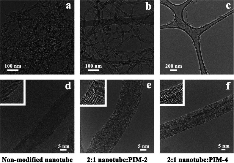
Fig. 4. Transmission electron microscopy images for (panel (a)) pristine multi-walled carbon nanotubes, (panel (b)) PIM-2-modified multi-walled carbon nanotubes, and (panel (c)) PIM-4-modified multi-walled carbon nanotubes. Panel (d) high-resolution transmission electron microscopy for pristine multi-walled carbon nanotubes; the magnified area in the box shows the smooth surface of the walls of the carbon nanotube. Panel (e) high-resolution transmission electron microscopy for the PIM-2-modified multi-walled carbon nanotubes; the magnified area in the box shows the walls of the carbon nanotube with a thin layer of the coating polymer on the surface. Panel (f) high-resolution transmission electron microscopy for the PIM-4-modified multi-walled carbon nanotubes; similar to panel (e), the magnified area in the box shows the walls of the carbon nanotube with a thin layer of the coating polymer on the surface. The information about the chemical structures of conjugated polyelectrolytes PIM-2 and PIM-4 is described in ref. 78. The figure is adapted with permission from ref. 78. Copyright 2015, American Chemical Society.

Fig. 4