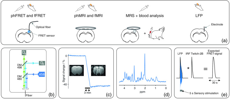
Fig. 1.
(a) Study design: functional () and pharmacological (ph) experiments were performed using fiber-based FRET recordings, MRI, MRS, blood analysis, or LFP recordings. For FRET recordings, a fiber was implanted in the S1Fl of Twitch-2B- or Laconic-expressing animals. For MRI and MRS, a 2- and 1-cm surface coil were used, respectively. A cortical voxel was investigated using MRS. For blood analysis, blood was sampled from the tail vein. LFP signals were recorded as difference between the electric currents of S1Fl and cerebellum using implanted electrodes. (b) Schematic of fiber-based FRET setup: a laser (458 nm) emitted excitation light (dark blue). Excitation light was reflected by a dichroic mirror (CWL: 480 nm) and coupled into a fiber. Fluorescent light emitted by the FRET sensors was separated using a second dichroic mirror (CWL: 498 nm): Fluorescent light emitted by donor fluorophores (light blue) was reflected and detected by an APD (). Light emitted from acceptor fluorophores (green) transmitted to the mirror and was detected by a second APD (). (c) Exemplary MRI measurement during contrast agent injection: signal decreases roughly 40% altering contrast notably. (d) Exemplary in vivo NMR spectrum. (e) Calculation of expected FRET ratios of Twitch-2B-expressing animals: Envelope of LFP response (blue) to sensory stimulation was convolved with the IRF of Twitch-2B.39

