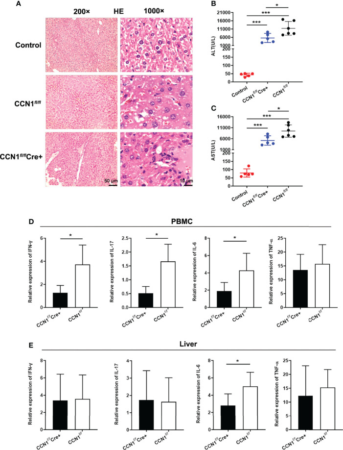
Figure 5.
Knocking down CCN1 expression attenuated liver injury and inflammation in ConA induced hepatitis mice. (A) Representative H&E staining images of liver tissues from ConA induced hepatitis mice. Original magnification ×200 (left, upper, scale bar: 50 µm), ×1000 (right, scale bar: 10 µm). (B) Serum levels of both ALT and AST in CCN1 fl/fl Cre+ (blue circle), CCN1 fl/fl (red circle) and control (black circle) mice. (C) mRNA expression of inflammatory cytokines IFN-γ, IL-17, IL-6 and TNF-α in PBMC from CCN1 fl/fl Cre+ and CCN1 fl/fl mice. (D) mRNA expression of IFN-γ, IL-17, IL-6 and TNF-α in PBMC from CCN1 fl/fl Cre+ and CCN1 fl/fl mice. (E) mRNA expression of IFN-γ, IL-17, IL-6 and TNF-α in liver from CCN1 fl/fl Cre+ and CCN1 fl/fl mice. *p<0.05, ***p<0.001.

