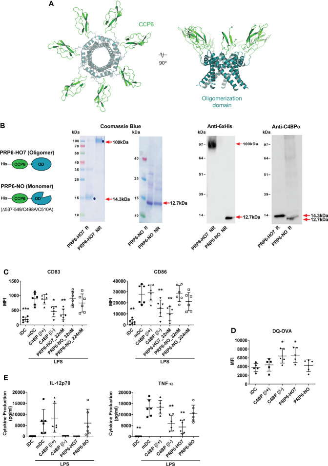
Figure 3.
Oligomerization is necessary to preserve the immunomodulatory activity of the C4BP α-chain CCP6 domain. (A) Molecular modeling of PRP6-HO7. PRP6-HO7 homo-oligomer structure prediction by comparative protein structure modeling with MODELLER. PRP6-HO7 is shown in cartoon representation. The N-terminal CCP6 domain and the C-terminal oligomerization domain are shown in green and blue, respectively. (B) Schematic structure of the monomer chain from the PRP6-HO7 heptamer, and from PRP6-NO, unable to oligomerize because of a mutated/truncated OD. PRP6-HO7 and PRP6-NO were visualized by SDS-PAGE and Coomassie Blue staining, and by Western analysis against anti-His and anti-C4BP α-chain antibodies, under both reducing (R) (PRP6-HO7 monomer, 14.3 kDa; PRP6-NO, 12.7 kDa) and non-reducing (NR) (PRP6-HO7 oligomer, 100 kDa; PRP6-NO, 12.7 kDa) conditions. Red arrows indicate the respective molecular weights. (C) Human Mo-DCs were incubated throughout their differentiation process with C4BP(β+), C4BP(β-) (both at 12 nM) and the variants PRP6-HO7 and PRP6-NO (both at 32 nM, unless otherwise stated). DC maturation was achieved by LPS treatment. Cells were then collected, washed, and analyzed by flow cytometry for cell surface expression of the activation marker CD83 and the co-stimulatory molecule CD86. MFI, median fluorescence intensity for the different surface markers. The results shown are the mean ± SD from 6 independent donors (*p < 0.05; **p < 0.01; ***p < 0.001 compared with mDC). (D) Comparative endocytic activity of Mo-DCs was also assessed by flow cytometry, measuring fluorescent DQ-OVA internalization (receptor-mediated endocytosis) at the differentiation stage, after treatment with C4BP(β+), C4BP(β-), PRP6-HO7, and PRP6-NO. Data shown are the mean MFI ± SD from 5 independent experiments (*p < 0.05 compared with iDC). (E) The concentrations of IL-12p70 and TNF-α were analyzed in the cell supernatants from (C), except the PRP6-NO_224 nM sample, by ELISA. iDC, untreated, immature DCs; mDC, untreated, LPS-matured DCs. The results shown are the mean ± SD from 6 independent donors performed in duplicate (**p < 0.01 compared with mDC).

