
Figure 5.
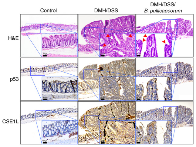
Representative immunohistochemical staining images for the expression of p53 and CSE1L proteins in the colon tissues. Colon tissues were sectioned from the following different groups: Control group, consisting of mice that did not receive any chemical treatment or B. pullicaecorum admin-istration; DMH/DSS group, consisting of mice that received DMH through intraperitoneal injection and DSS in their drinking water but did not receive B. pullicaecorum; and DMH/DSS/B. pullicaecorum group, consisting of mice that received DMH/DSS and B. pullicaecorum. Colon tumors exhibited weak nuclear staining of p53 and markedly increased expression of CSE1L. Insets show the magnified views of the boxed areas. Red arrows indicate the intramucosal adenocarcinoma in DMH/DSS group (middle panel) and the low-grade adenoma in DMH/DSS/B. pullicaecorum group (right panel). Scale bar, 50 µm for inset. CSE1L, chromosome segregation 1-like protein; DMH, 1,2-dimethylhydrazine; DSS, dextran sulphate sodium; B. pullicaecorum, Butyricicoccus pullicaecorum.