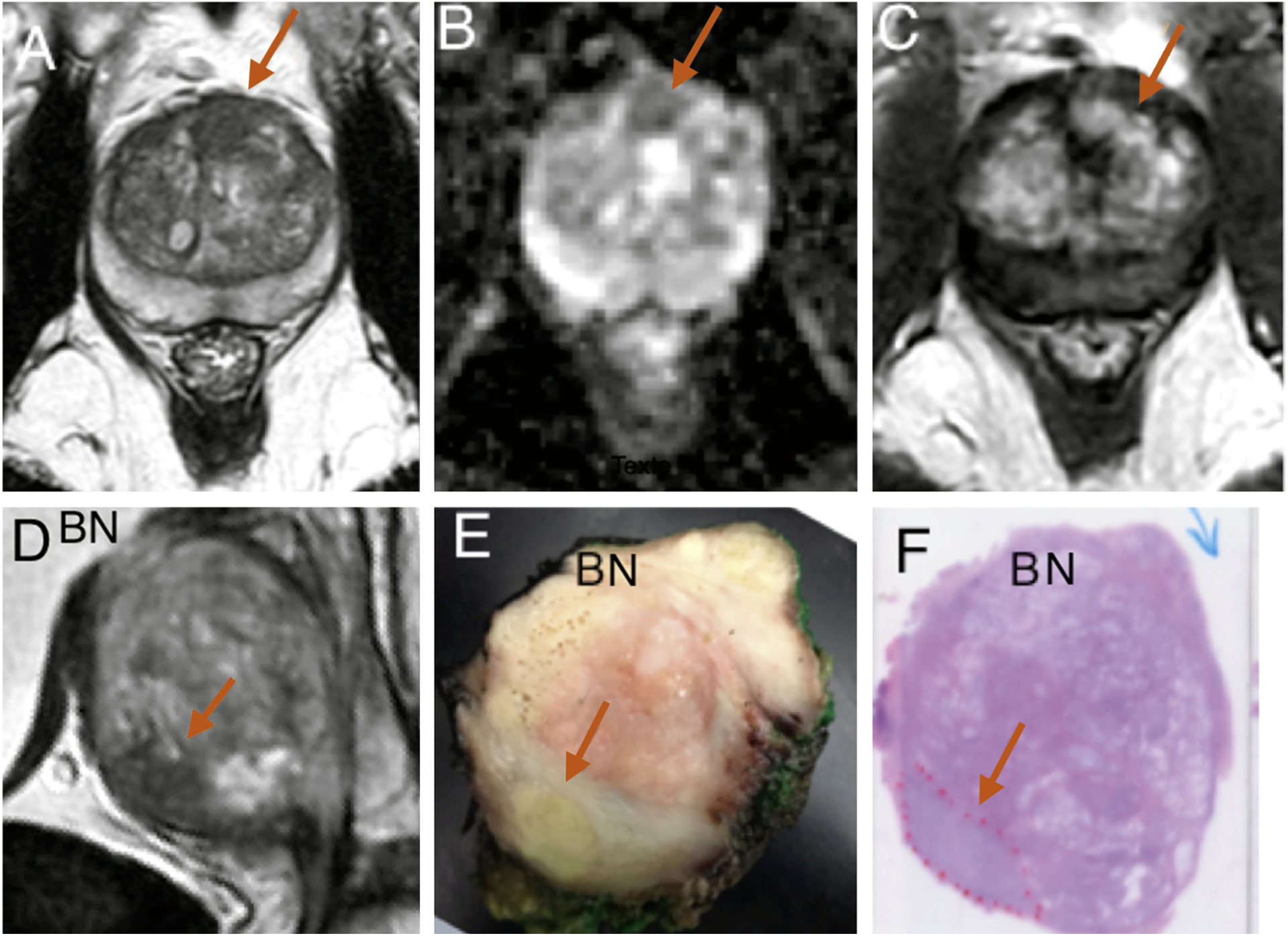
Fig. 2 –

Case 17 (prostate-specific antigen [PSA] 7.13 ng/ml; prostate volume 73 cm3]. Isolated anterior 2.4-cm3 lesion suspicious at magnetic resonance imaging (MRI) in the anterior fibromuscular stroma on the midline and anterior left transition zone lobe (arrows). (a) MRI transverse T2. (b) MRI transverse apparent diffusion coefficient map. (c) MRI transverse dynamic contrast-enhanced sequences. Targeted biopsies were positive for 6-mm Gleason score (GS) 6 (3 + 3) cancer. (d) MRI T2 parasagittal view showed anterior cancer area at the anterior and inferior aspect (arrow). (e) Fixed midsagittal section showed yellow area suspected of malignancy at the anterior and inferior aspect (arrow). (f) Hematoxylin and eosin whole-mount sagittal histologic section confirmed cancer (red dotted line) area of 4 cm3, GS 7 (4 + 3), pT2, R0, and postoperative PSA of 0.4 ng/ml at 3 mo.
BN = bladder neck.
