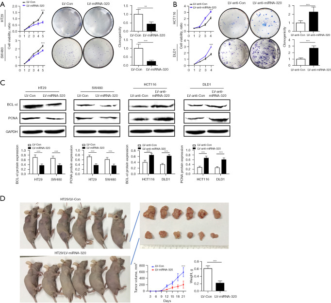
Figure 3.
miRNA-320 inhibits cell proliferation. (A,B) Cell proliferation was measured by CCK-8 assays and plate colony formation assays (stained with Giemsa) in HT29 and SW480 cells transfected with LV-miRNA-320 or control (A) and HCT116 and DLD1 cells transfected with LV-anti-miRNA-320 or control (B). (C) The protein expression of BCL-xl and PCNA in HT29, SW480 cells, HCT116, and DLD1 cells. The same column was GAPDH as an internal control. (D) Image of subcutaneous xenograft tumors. Nude mice were injected with 1×10⁷ HT29 cells transfected with LV-miRNA-320 (n=6/group). The tumors were extracted after 21 days. Analysis of the tumor volume measured every 3 days. Tumor weight in each group at the end of the experiment. Data are expressed as the mean ± SD. **, P<0.01; ***, P<0.001. miRNA, microRNA; CCK-8, Cell Counting Kit-8; BCL-xl, B-cell lymphoma-extra large; LV, lentiviral vector; PCNA, proliferating cell nuclear antigen; GAPDH, glyseraldehyde-3-phosphate dehydrogenase; SD, standard deviation.

