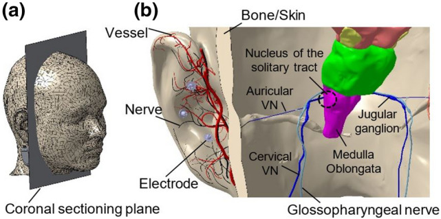
Fig. 1.

a Location of the coronal sectioning plane of the head to show the relevant VN network in b. The posterior side of the sectioned head with three needle electrodes in the cymba conchae region of the external ear, which stimulate aVN endings. The stimulation is projected by the aVN branch extension from the external ear to the vagus nerve (through external auditory meatus) into the jugular ganglion and eventually to the nucleus of the solitary tract. Vessels and nerves are shown in red and black, respectively. External ear model with the associated vessels and nerves are taken from [14, 15], the head model from [16]
