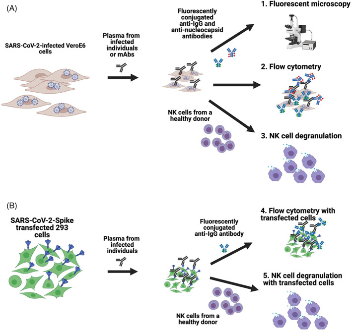
FIGURE 1.

Schematics of the assays developed to assess antibody recognition of SARS‐CoV‐2 on the surface of infected and transfected cells. Vero E6 cells were infected with SARS‐CoV‐2 and then incubated with plasma from infected individuals or anti‐SARS‐CoV‐2 spike‐specific monoclonal antibodies. Cells were then either stained with fluorescently conjugated anti‐IgG and anti‐Nucleocapsid antibodies to assess using fluorescent microscopy or flow cytometry, or incubated with NK cells from a healthy human donor to measure the capacity of antibodies to mediate NK cell degranulation (A). For transfected cell assays, 293 cells were transfected with SARS‐CoV‐2 spike plasmid, incubated with plasma from infected individuals and then stained with fluorescently conjugated anti‐IgG to assess using fluorescent microscopy or flow cytometry, or incubated with NK cells from a healthy human donor to measure the capacity of antibodies to mediate NK cell degranulation (B) [Color figure can be viewed at wileyonlinelibrary.com]
