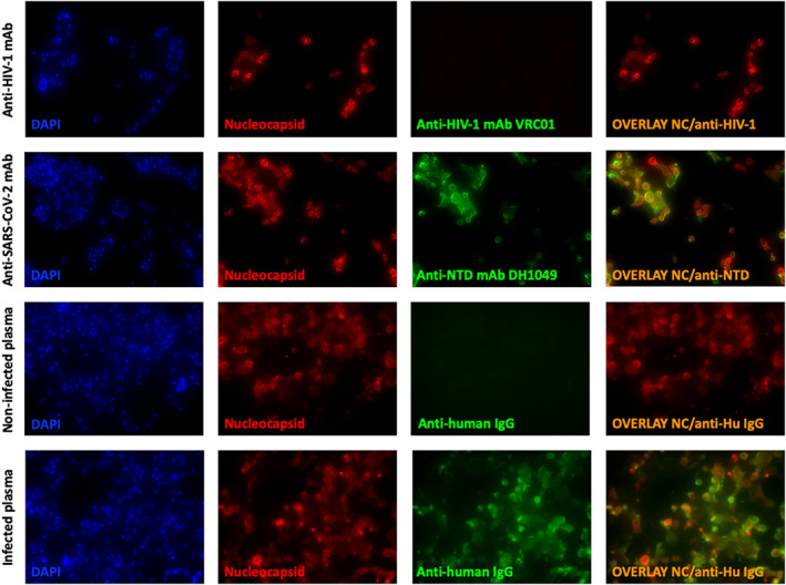
FIGURE 2.

Fluorescent microscopy showing antibody binding SARS‐CoV‐2‐infected cells. Infected Vero E6 cells were stained intracellularly with an anti‐SARS‐CoV‐2 Nucleocapsid antibody (red), on the surface with controls (anti‐HIV‐1 monoclonal antibody, VRC01, or plasma from an uninfected individual) or SARS‐CoV‐2 specific antibody (anti‐SARS‐CoV‐2 NTD mAb, DH1049, or sera from a SARS‐CoV‐2‐infected individual) (green). Nucleated cells were identified using DAPI (blue) and an overlay of anti‐SARS‐CoV‐2 Nucleocapsid and anti‐SARS‐CoV‐2 antibodies bound to the surface of cells is shown (right column). Cells were visualized and pictures taken at 100× [Color figure can be viewed at wileyonlinelibrary.com]
