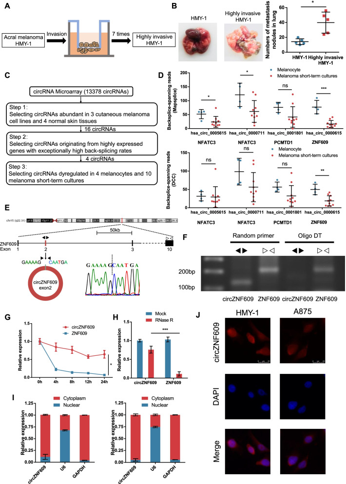
Fig. 1.
The identification and characteristics of circZNF609 in human melanoma cells. a The sketch map of cell invasion model. b In vivo lung metastasis model to verify the different metastatic potential between HMY-1 and highly invasive HMY-1 sublines. c Flowchart illustrating the screening criteria of potential regulatory circRNAs in acral melanoma and cutaneous melanoma. d The expression of 4 circRNAs in 4 melanocytes and 10 melanoma short-term cultures of GSE138711 according to Mapsplice algorithm (top) and DCC algorithm (bottom). e The formation of circZNF609. circZNF609 derived from back-spliced exons 2 of genomic ZNF609. The existence of circZNF609 was detected by Sanger sequencing. Black arrows represent divergent primers and white arrows represent convergent primers. f PCR products with different primers showing circularization of circZNF609. Random primers amplified circZNF609 in cDNA. Oligo DT did not amplify circZNF609 in cDNA. Linear ZNF609 was used as a control. g qRT–PCR analysis for the expression of circZNF609 and ZNF609 mRNAs after treatment with Actinomycin D in HMY-1 cells. h qRT–PCR analysis for the expression of circZNF609 and ZNF609 mRNAs after treatment with RNase R in HMY-1 cells. i Nucleoplasmic separation assay showing that circZNF609 mainly located in the cytoplasm in HMY-1 (left) and A875 cells (right). GAPDH and U6 were applied as positive controls in the cytoplasm and nucleus, respectively. j FISH assays showing the predominant cytoplasmic distribution of circZNF609 in HMY-1 and A875 cells

