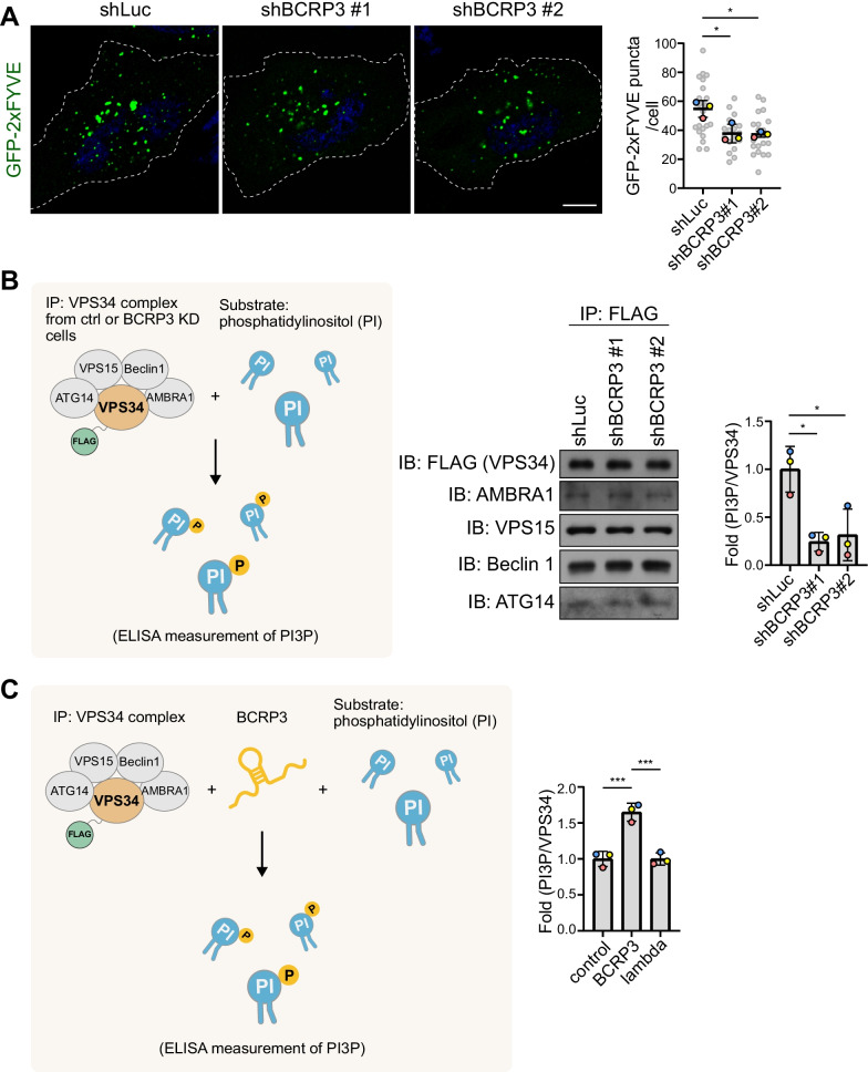
Fig. 5.
BCRP3 stimulates the activity of VPS34 complex. A Confocal microscopy analysis of control or BCRP3-deficient HeLa cells, transiently transfected with GFP-2xFYVE and starved in EBSS for 1 h. Representative images are shown on the left and quantitative data are on the right. Bar, 10 μm. Data are means ± SD from three independent experiments and 7 cells per group per experiment were counted. P values are determined by one-way ANOVA with Tukey’s post hoc test, *P < 0.05. B FLAG-VPS34 was immunoprecipitated from control or BCRP3-deficient HeLa cells. The immunocomplexes were analyzed by Western blot (middle) or incubated with phosphatidylinositol (PI) substrate and ATP (left). PI3P production was measured by ELISA assay and normalized to FLAG-VPS34 protein levels (right). C FLAG-VPS34 immunoprecipitated from transfected HeLa cells was incubated with PI substrate and ATP, together with in vitro transcribed BCRP3 or lambda RNA (left). PI3P production was measured by ELISA and normalized to FLAG-VPS34 protein levels (right). Data in (B) and (C) are means ± SD from three independent experiments. P values are determined by one-way ANOVA with Tukey’s post hoc test, *P < 0.05, ***P < 0.001

