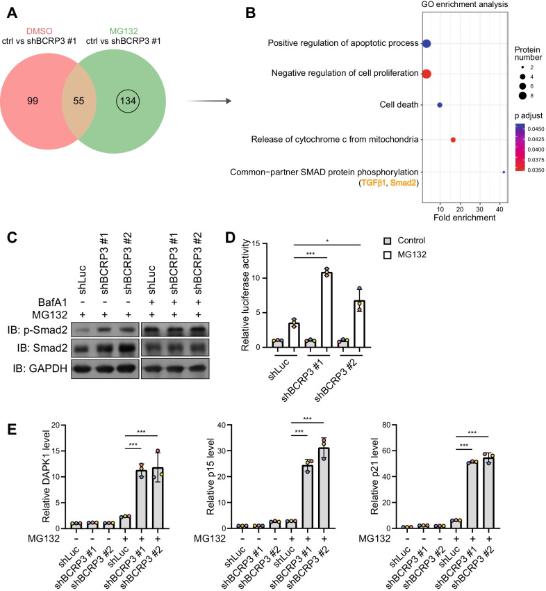
Fig. 7.
BCRP3 deficiency in proteotoxicity leads to the accumulation of proteins involving in growth inhibition, cell death, and TGF-β/Smad2 signaling. A Venn diagram showing the numbers of enriched proteins after BCRP3 knockdown together with or without 10 µM MG132 treatment for 12 h. B GO enrichment analysis of the 134 proteins shown in (A). Selective enriched GO terms are shown by the order of fold enrichment (bottom to top). C Western blot analysis of indicated proteins in control or BCRP3-deficient HeLa cells treated with 10 µM MG132 together for 12 h together with or without 200 nM bafilomycin A1 for 2 h. D Control or BCRP3-deficient HeLa cells were transfected with 4 × SBE-Luc reporter construct, treated with 10 µM MG132 for 12 h and analyzed for luciferase activity. E qRT-PCR analysis of relative DAPK1, p15, and p21 levels in control or BCRP3-deficient HeLa cells treated with 10 µM MG132 for 12 h. Data in (D), (E) are means ± SD from three independent experiments. P values are determined by one-way ANOVA with Tukey’s post hoc test, *P < 0.05, ***P < 0.001

