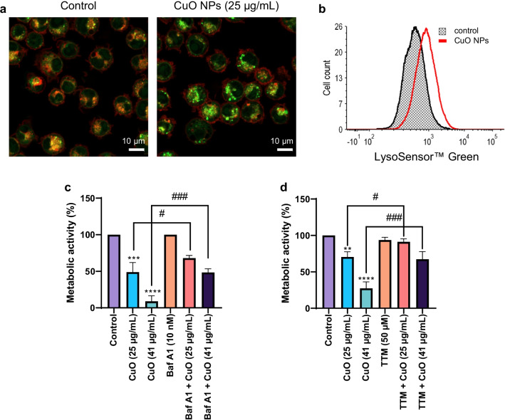
Fig. 3.
CuO NPs trigger lysosomal acidification. a RAW264.7 macrophages were labeled using LysoSensor™ Green following exposure to CuO NPs (25 µg/mL) for 6 h and visualized by confocal microscopy. Scale bars: 10 µm. b LysoSensor™ Green fluorescence in control cells and cells exposed to CuO NPs (25 µg/mL) was determined by flow cytometry. Preincubation with bafilomycin A1 (10 nM) (c) and TTM (50 µM) (d) protected the RAW264.7 macrophages from CuO NP-triggered cell death as shown using the Alamar blue assay. Data are mean values ± S.D. (n = 3). **p < 0.01; ***p < 0.001; ****p < 0.0001 (significant difference between control and treatments); #p < 0.05; ###p < 0.001 (significant difference between treatments)

