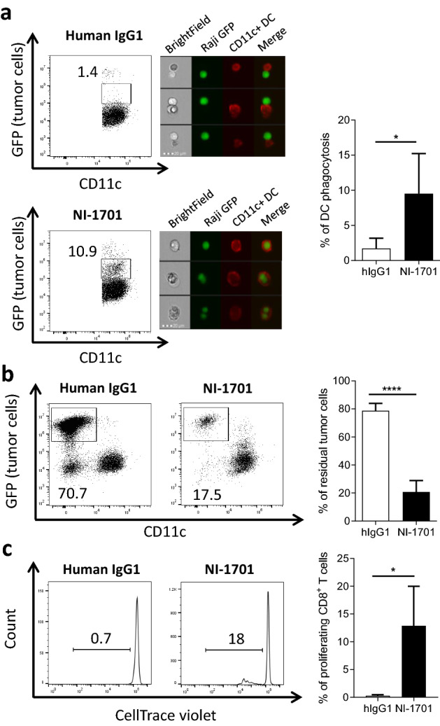
Fig. 4.

NI-1701 induces tumor cell phagocytosis by bone-marrow-derived dendritic cells and promotes cross-priming of CD8+ T cells. Bone marrow-derived mouse dendritic cells (CD11c+) from BALB/c mice were cocultured for 2 h 30 (a) or 24 h (b) with Raji GFPhi tumor cells (1:1 ratio) in the presence of hIgG1 or NI-1701. Representative flow cytometry plots for hIgG1 and NI-1701 treated groups are depicted, and the percentage of phagocytosis is indicated (left panel). Phagocytic events were confirmed by imaging flow cytometry acquisition in the CD11c+GFP+ gate with tumor cells in green fluorescence and CD11c+ dendritic cells in red fluorescence (middle panel). The mean percentage of phagocytosis at 2 h 30 ± SD is shown (right panel), n = 5 independent experiments. b Percentage of residual Raji GFPhi tumor cells after 24 h of coculture with CD11c+ DCs was determined by the analysis of remaining GFP+ tumor cells within the total live cell gate. n = 5 experiments. c Bone-marrow derived dendritic cells from BALB/c mice were cocultured overnight with hemagglutinin-expressing Raji tumor cells (Raji HA-GFP) in the presence of human IgG1 or NI-1701 (ratio 1:1). The next day, HA-specific CD8+ T cells from CL-4 transgenic mice (bearing HA-specific TCR on CD8+ T cells) labelled with CellTrace violet were added (ratio 1:5). Analysis of CD8+ T cell proliferation was performed 3 days later. Plots represent an illustration of proliferating CD8+ T cells (left panel). Mean ± SD was calculated from 4 independent experiments (right panel). Significance was determined by unpaired t test. *p < 0.05, ****p < 0.0001
