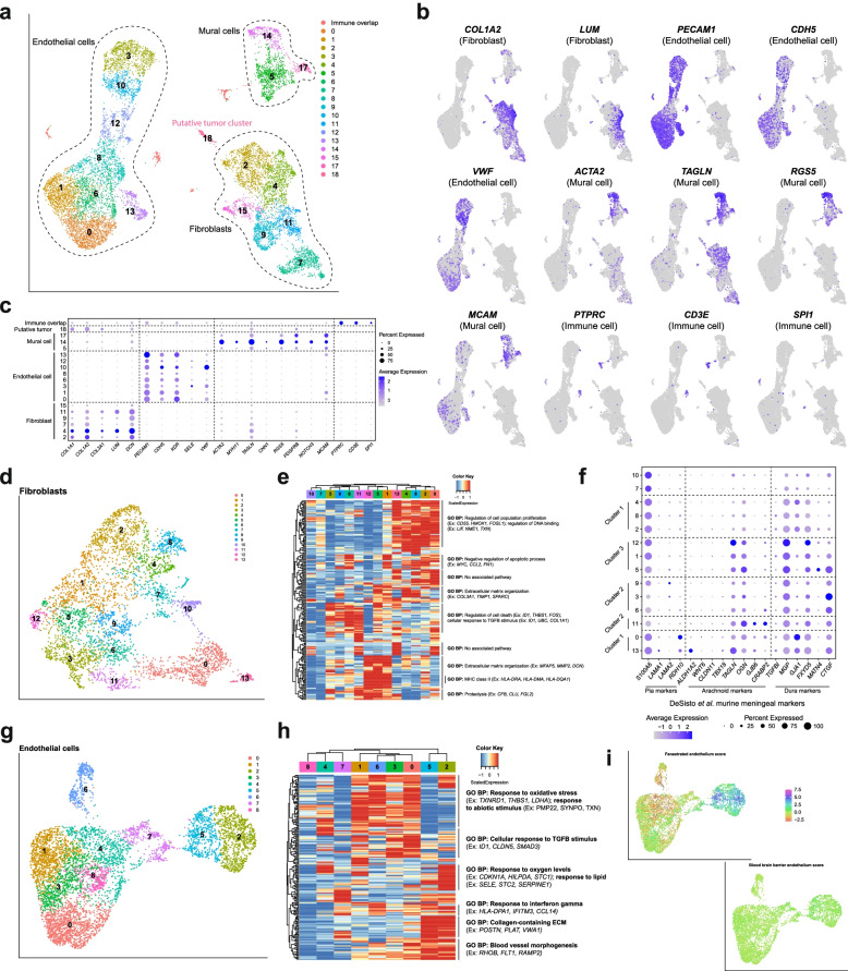
Fig. 3.
Functionally diverse non-immune cells comprise a significant proportion of human dura. A UMAP visualization of non-immune cells identified by cell type. B UMAP visualization of select endothelial, mural, and fibroblast markers. C Dot plot of representative gene expression of select cell type gene markers. D UMAP visualization of fibroblast endothelial cells. E Expression heatmap of top 15 genes of top 15 principal components of dura fibroblast cells with hierarchical clustering and associated functional enrichment analysis of gene clusters. F Dot plot of select meningeal fibroblast markers from Desisto et al. [39]. G UMAP visualization of dura endothelial cells. H Expression heatmap of top 15 genes of top 15 principal components of dura endothelial cells with hierarchical clustering and associated functional enrichment analysis of gene clusters. I UMAP visualization of fenestrated endothelium and blood-brain barrier scores

