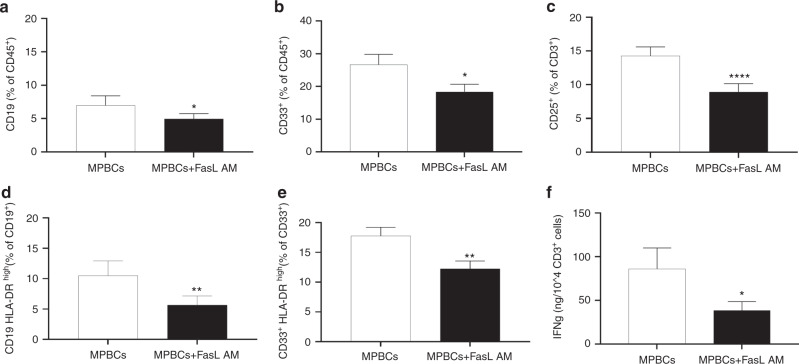
Fig. 3. FasL AM treatment of MPBCs decreases B cells, myeloid cells and activated T cells populations, reducing the antigen presentation potential and T cell activation potential.
MPBCs graft characterization following 100 ng/ml FasL AM treatment: (a) Percentage of B-lymphocytes (CD19+), (b) Myeloid cells (CD33+) and (c) Activated T cells (CD25+) per total CD45+ population. The percentage of HLA-DRhigh expressing on B-lymphocytes (d) and on myeloid cells (e) as marker for antigen presentation potential per total CD19+ or CD33+ population. N = 7, *P ≤ 0.05, **P ≤ 0.01, ****P ≤ 0.0001; Paired T-test. f Concentration of IFN-γ secreted to the medium, measured 24 h post T cell specific and normalized per seeded CD3+ cell number in culture. N = 8, *P ≤ 0.05; Paired T-test.

