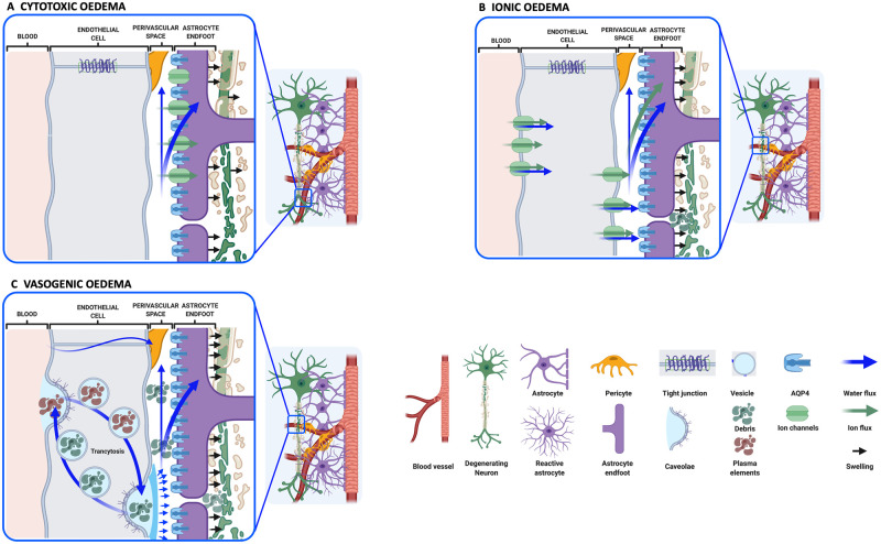
Figure 4.
Classification of CNS oedema. (A) Cytotoxic oedema is defined by astrocyte swelling (black arrows) followed by neuronal dendrite swelling. The net entry of water (blue arrows), most likely from the perivascular space, is caused by disruption of cellular ion homeostasis (green arrows) following hypoxic insult. (B) Ionic oedema is characterized by transcapillary sodium ion and anion fluxes associated with cellular uptake of ions from the perivascular CSF, and entry of water into the brain parenchyma. Astrocytes continue to be swollen (black arrows) by water from the perivascular space and the vascular compartment. Neuronal death produces cellular debris in the extracellular space (ECS). (C) Vasogenic oedema is a result of BBB dysfunction, possibly following ionic oedema. Increased transcytosis may contribute to the entry of plasma elements (brown), followed by water. Clearance of debris from the ECS produced by neuronal cell death may also occur by transcytosis (green). In some severe cases, the tight junctions between the endothelial cells are weakened leading to increased permeability of cerebral blood vessels to plasma components. Created using www.biorender.com.

