Figure 1.

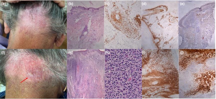
Clinical presentation and histology before (a–e) and after (f–j) the vaccine. (a) Purple patch on the occipital area, (b) H&E ×20: Lymphoid infiltration of the dermis with prominent folliculotropism, (c) CD3 ×40: CD3 expression, (d) CD4 ×20: CD4 expression, (e) CD30 ×20: Sparse CD30 positivity, (f) Lichenoid induration and small nodules on the periphery of the patch, (g) H&E ×20: dense, diffuse, full thickness lymphoid infiltration of the dermis, (h) H&E ×400: increased numbers of large neoplastic T lymphocytes among the lymphocytic population, (i) CD3 X 20: CD3 expression, (j) CD30 ×40: extensive CD30 positivity mainly in the large anaplastic T lymphocytes (transformation to peripheral CD30+ T‐cell lymphoma).
