
Figure 1.
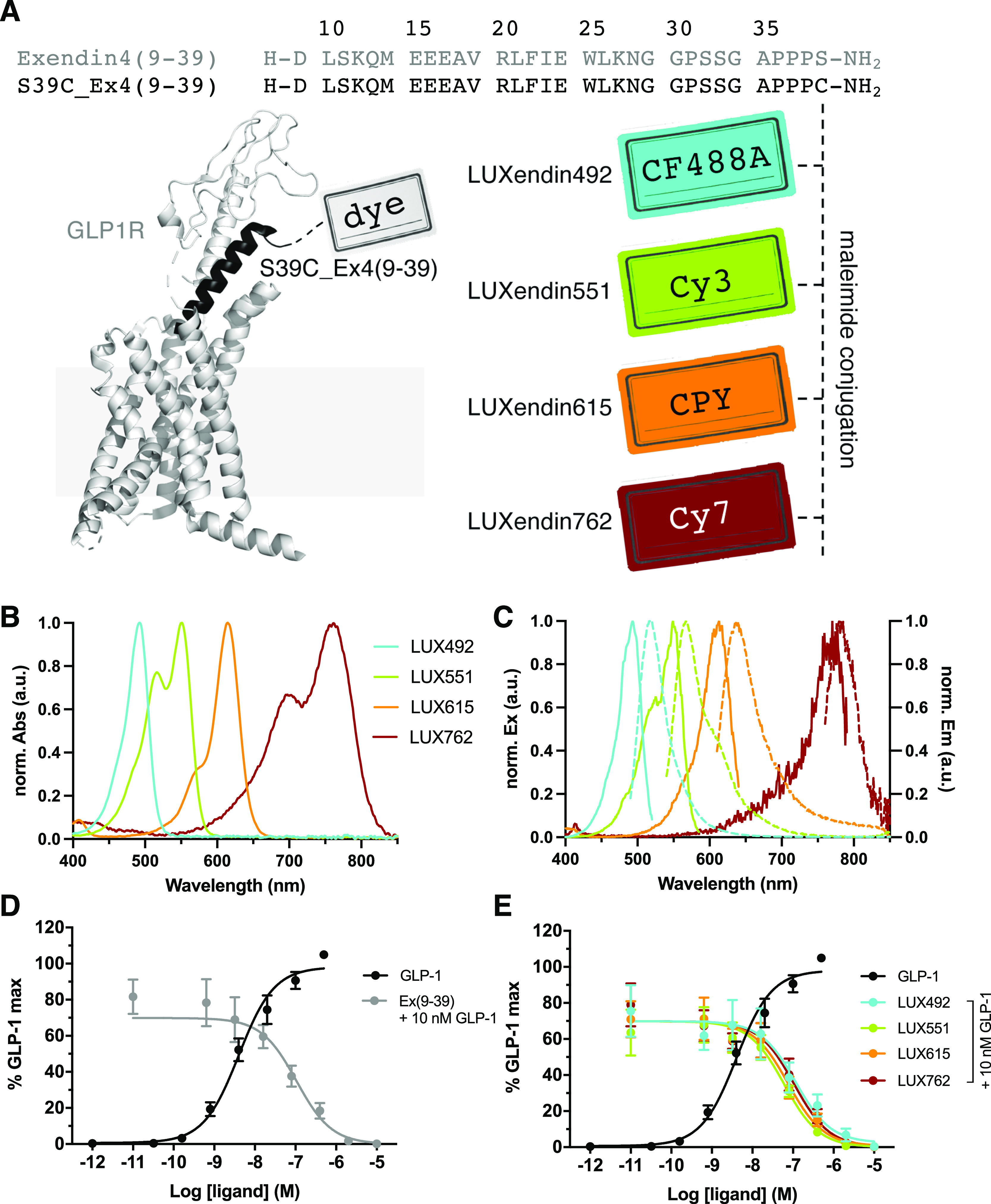
Sequence, structure, photophysical properties, and pharmacology of LUXendin492, LUXendin551, LUXendin615, and LUXendin762. (A) LUXendins are based on the antagonist Exendin4(9–39) with a S39C mutation to install fluorophores via late-stage thiol–maleimide chemistry. The model shows GLP1R in complex with a peptide ligand [pdb: 5VAI, cartoon obtained by the in-built building capability of PyMOL (Palo Alto, CA, USA)]. CF488A, Cy3, CPY, and Cy7 were installed as fluorescent labels to give LUXendin492, LUXendin551, LUXendin615, and LUXendin762, respectively. (B) UV/vis spectra of novel LUXendins. (C) Fluorescence excitation and emission spectra of LUXendins. (D) cAMP response in GLP1R-transfected HEK293 cells for GLP-1 (agonist, black) and Ex(9–39) (antagonist) in the presence of 10 nM GLP-1 (gray) (n = 6 independent repeats). (E) Sames as (D), but in response to LUXendins (colored), showing the antagonistic nature of the probes.