. 2018 Oct 29;8(64):36422–36429. doi: 10.1039/c8ra02418a

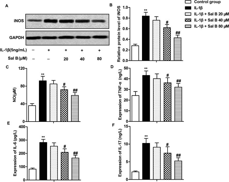
Fig. 2. Sal B inhibits the production of inflammatory mediators. IL-1β (5 ng mL−1) stimulated chondrocytes for 6 h in vitro and divided them into 5 groups. The control group, control cells + IL-1β induced group, IL-1β + Sal B 20 μM group, IL-1β + Sal B 40 μM group and IL-1β + Sal B 80 μM group. Adding the same amount of solvent were added into the control group and control cells + IL-1β induction group. (A and B) The expression of iNOS was detected by western blot; (C) the production of NO was measured by nitrate reductase. (D) mRNA levels of TNF-alpha in supernatant were detected by qRT-PCR. (E) mRNA levels of IL-6 in supernatant were detected by qRT-PCR. (F) mRNA levels of IL-17 in supernatant were detected by qRT-PCR. The GAPDH was used as the internal reference. **P < 0.01 versus control group, #P < 0.05, ##P < 0.01 versus IL-1β induction group.

Fig. 2