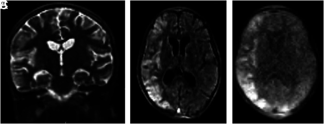
FIG 2.
Hyperfine Swoop MR images of a 6-year-old child who was admitted to our institution under the research study arm with a decreased level of consciousness and malaria parasitemia. The child’s MR imaging demonstrates typical findings associated with malarial encephalopathy of diffuse right-sided parietotemporal T2-weighted hyperintensity in the gray and subcortical white matter with an associated localized mass effect in the form of sulcal effacement on the coronal T2-weighted sequence (A), with corresponding restriction of diffusion on the DWI b=900 image and ADC images (B and C).

