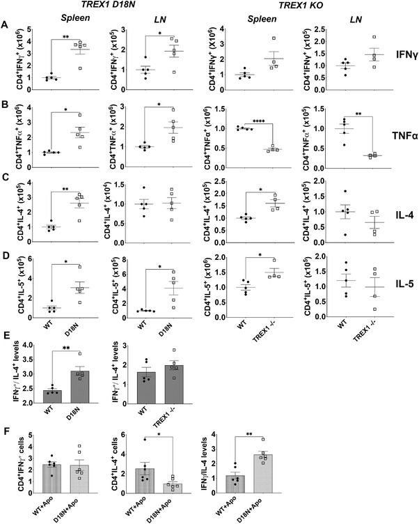
Figure 2.

Status of T‐helper cytokines in TREX1 deficiency. Spleen and lymph nodes (LN) from female TREX1 D18N and TREX1 knockout mice were stimulated with PMA/Ionomycin for 5 h and levels of Th1 cytokines IFN‐γ (A), TNF‐α (B), and Th2 cytokines IL‐4 (C) and IL‐5 (D) were analyzed by flow cytometry. (E) The ratio of IFN‐γ to IL‐4 producing CD4 T cells indicated a significant Th1 bias as shown in the spleen of TREX1 D18N mice, but did not reach statistical significance in TREX1 KO animals. (F) In vitro T‐cell stimulation assay using BM‐derived macrophages as described in the Materials and Methods showed that TREX D18N macrophages induced similar levels of %IFN‐γ producing T cells, but much fewer %IL‐4 producing cells. A ratio of IFN‐γ/IL‐4 confirmed the greater Th1 bias induced by the TREX1 D18N macrophages. Data presented as mean ± SEM relative to WT mice (normalized absolute numbers) and a total of four to five mice per group over three independent experiments, with the dots representing individual mice, and three independent experiments for in vitro assays. *p < 0.01, **p < 0.001 as calculated by two‐tailed unpaired Student's t‐test. Also see Supporting information Fig. S2 for data in percentages.
