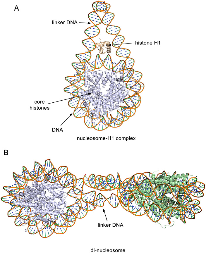
Figure 1.
Crystal structures of the nucleosome. a) The nucleosome in complex with histone H1. Octamer of histones H2A, H2B, H3 and H4 as well as DNA, wrapped around the histone core, are shown in a ribbon diagram and colored light blue and orange, respectively. Histone H1 is shown in wheat. PDB ID: 5nl0. b) Di-nucleosome with coloring the same as in (a) for one nucleosome with the octamer shown in green for the second nucleosome. PDB ID: 1ZBB.

