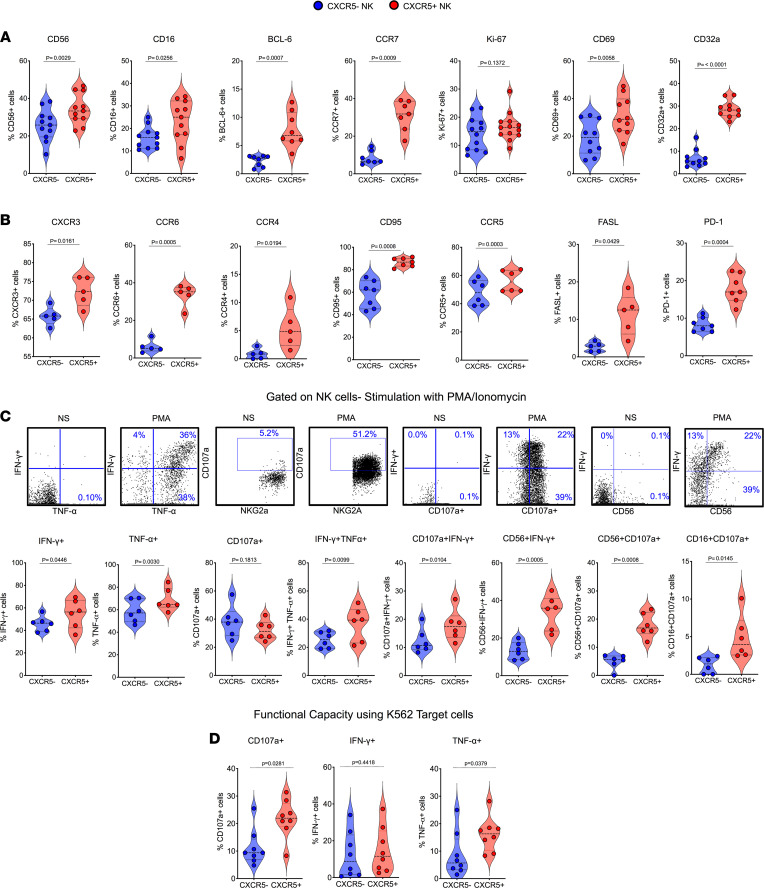
Figure 2. Follicular CXCR5+ NK cells display activated phenotype and heightened functionality.
(A) Violin plots showing the expression of different indicated markers (CD56, CD16, BCL-6, CCR7, Ki-67, CD69, CD32a) on CXCR5+ and CXCR5– NK cells; data shown for week 14 after SHIV infection (n = 11). (B) Expression of chemokine receptors (CXCR3, CCR6, CCR4) and CD95, CCR5, FASL, PD-1 on CXCR5+ and CXCR5– NK cells (n = 6). (C) Cytokine expression and degranulation (CD107a+ staining) profiles of CXCR5+ and CXCR5– NK cells after 6 hours of ex vivo culture in presence (PMA) or absence (NS) of PMA and ionomycin determined by intracellular cytokine staining and flow cytometry (n = 6). (D) Similar assessment of functionality of CXCR5+ and CXCR5– NK cells after 6 hours of coculture with MHC class I–devoid K562 target cells (n = 8). Wilcoxon’s matched-pairs signed rank test was used to compare the frequencies of CXCR5+ and CXCR5– NK cells in the lymph nodes.

