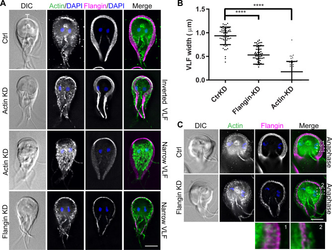
Fig 2. Flangin and GlActin are necessary for flange assembly.
(A) Trophozoites were stained for GlActin (green), Flangin-HA (magenta), and DNA (blue). Note that images are scaled to display the remaining protein and are not intended to show differences in protein levels. GlActin knockdown (KD) resulted in inverted and collapsed flange morphology. Flangin-HA KD similarly resulted in a thin flange phenotype. (B) Quantification of flange width when measured at the cell anterior from three independent experiments; control (n = 57), Flangin-HA (n = 54), and actin (n = 51). Statistical significance was evaluated for Flangin-KD and Actin-KD respectively, t test. ****, P<0.0001. (C) Mitotic Flangin depleted cells were capable of extending their flange beyond the leading edge of Flangin. The insets show actin beyond the leading edge of Flangin-HA in Flangin-HA KD cells during mitotic flange extension. Scale bars = 5 μm.

