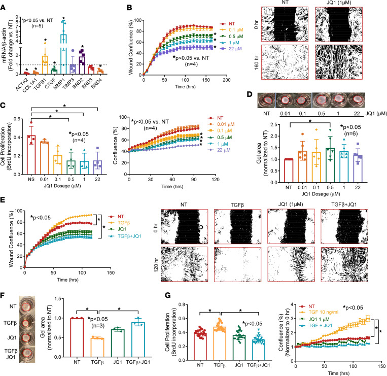
Figure 2. Inhibition of BETs shows potent antifibrotic properties in dcSSc fibroblasts.
(A) At 1 μM, JQ1 significantly downregulated ACTA2, COL1A1, CTGF, and BRD4 expression in dcSSc fibroblasts and upregulated MMP1, TGFB1, and BRD2. JQ1 did not affect TIMP1 and BRD3 expression. n = 5 patients. (B) Migration of dcSSc fibroblasts was significantly inhibited by JQ1. Wound confluence indicates the area occupied by cells that migrated into the wound gap. Representative pictures of JQ1 at 1 μM are shown. n = 4 patients. (C) Inhibition of BETs by JQ1 significantly reduced cell proliferation of dcSSc fibroblasts. Cell growth was analyzed by BrdU uptake in cells or monitored by Incucyte live-cell imaging system. n = 4 patients. (D) Gel contraction by dcSSc fibroblasts was inhibited by JQ1 at 0.5 μM. n = 6 patients. (E–G) Cell migration (n = 4), proliferation (n = 4), and gel contraction (n = 3) significantly increased after TGF-β treatment in normal dermal fibroblasts and can be inhibited by coincubation of 1 μM JQ1. Results are expressed as mean ± SD or mean ± SEM (time courses in B, C, E, and F); P < 0.05 was considered significant. Significance was determined by unpaired 2-tailed t test and Mann-Whitney U test (A), 2-way ANOVA (B, C, E, and G), 1-way ANOVA (F), and Kruskal-Wallis test (D and G). NT, no treatment.

