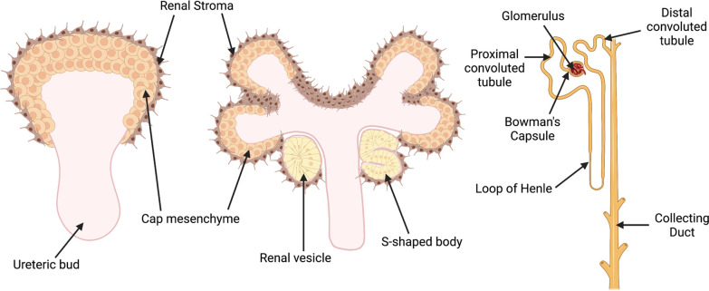
Figure 2. Schematic illustration of the stages of metanephric kidney development.
Signals from the ureteric bud trigger condensation of the metanephric mesenchyme to form a cap of nephron progenitors (cap mesenchyme) around the ureteric bud tips. The cap mesenchyme undergoes a mesenchymal-epithelial transition to form renal vesicles, which develop sequentially into comma- and S-shaped bodies. These structures connect to the ureteric bud stalk, which give rises to the collecting duct. Cells in the proximal domain of the S-shaped body differentiate into specialized epithelial cells of the mature renal corpuscle (i.e., podocytes and Bowman’s capsule cells), while cells in the mid- and distal portions differentiate into the tubular segments of nephron (proximal tubules, loops of Henle, and distal tubules). Created with BioRender.com.

