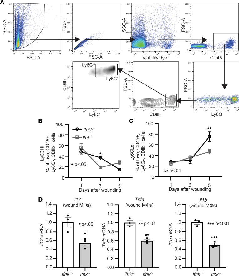
Figure 2. IFN-κ regulates macrophage inflammatory profile during normal tissue repair.
(A) IFN-κ–KO and control wound cell isolates were processed for flow cytometry using the following gating strategy selecting for single cells, live (viability dye–), CD45+, Ly6G−, CD11b+, Ly6Chi, or Ly6Clo. (B and C) Flow cytometry quantification of Ly6Chi and Ly6Clo cells in wounds (n = 4–6 per group). (D) Wound monocyte/macrophages (MΦ) (CD3–CD19–Ly6G–CD11b+) were isolated from WT and IFN-κ–KO mice on day 3; n = 3 per group, repeated in triplicate. Gene expression of inflammatory cytokines Tnf, Il1b, and Il12 was measured via qPCR. Data were analyzed for variances, and 2-tailed Student’s t test or 1-way ANOVA was performed. *P < 0.05, **P < 0.01, and ***P < 0.001. Data are presented as mean ± SEM.

