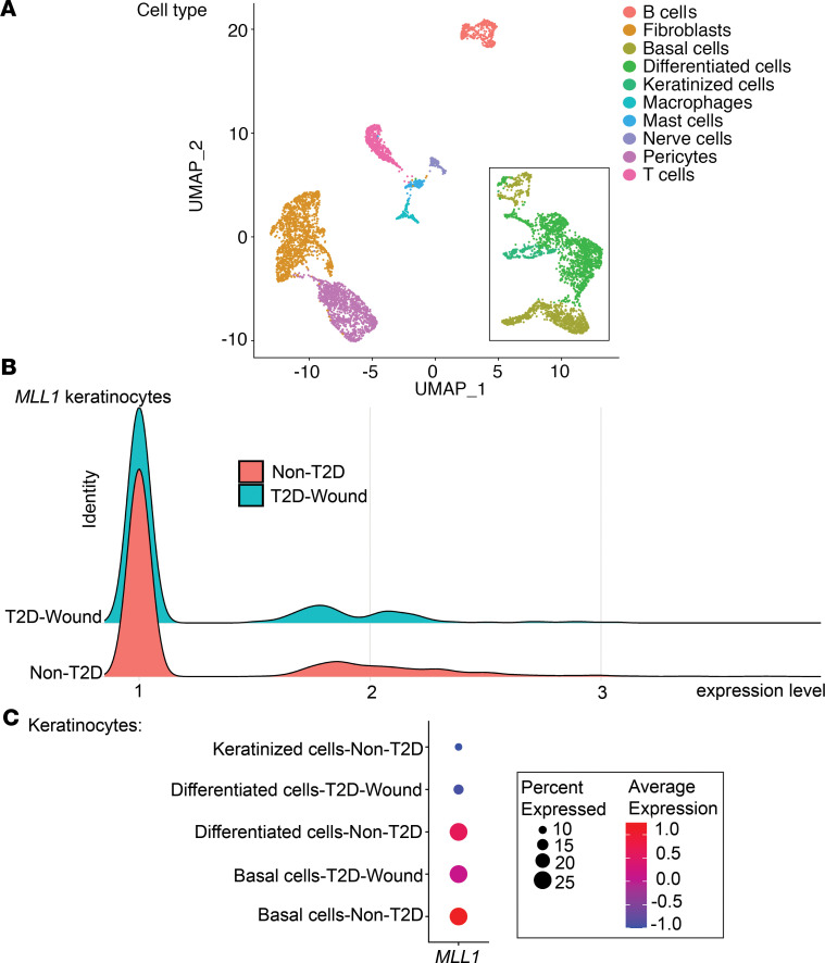
Figure 5. scRNA-Seq of Mll1 expression is decreased in human T2D wounds.
(A) Cluster analysis was performed using the UMAP technique of single-cell sequencingT2D (n = 1) and nondiabetic wound (n = 2) samples. The black box outlines the keratinocyte populations: basal cells, differentiated cells, keratinized cells. (B) Ridge plot demonstrating expression levels of MLL1 within keratinocytes in human T2D and non-T2D samples. (C) Dot plot demonstrating MLL1 expression within keratinocyte populations in human T2D and non-T2D samples. Dot size corresponds to the proportion of cells within the group expressing MLL1, and dot color corresponds to expression level.

