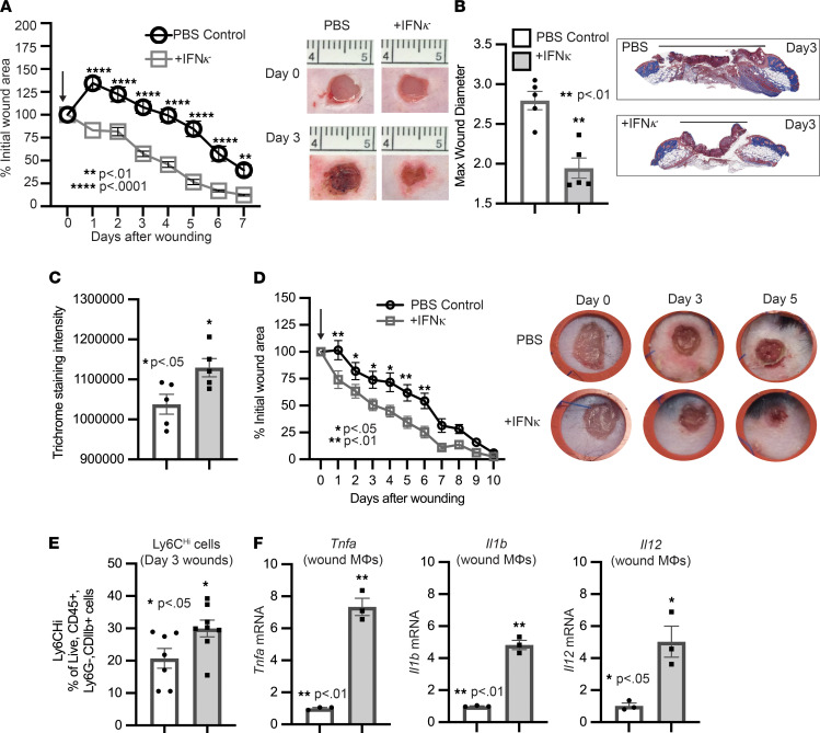
Figure 6. Administration of IFN-κ improves diabetic wound healing.
(A) The 4 mm punch biopsy wounds were created on DIO, and wounds were injected daily starting on day 0 for 3 days after injury with IFN-κ (0.5 μg/100 μL) or PBS control (2 wounds per mouse, n = 5 mice per group, repeated once). The arrow indicates when injections started. The change in wound area was recorded daily and analyzed with ImageJ software. Representative photographs of the wounds were taken on days 0 and 3. (B) Max wound diameter at day 3 for IFN-κ and PBS control; n = 5 per group. Trichrome staining representative picture. (C) Trichrome staining was calculated using ImageJ software (n = 5 mice/group). (D) The 4 mm punch biopsy wounds were created and splinted on DIO mice, and wounds were injected starting on day 0 for 3 days after injury with IFN-κ (0.5 μg/100 μL) or PBS control (1 wound per mouse, n = 7–8 mice per group). The arrow indicates when injections started. The change in wound area was recorded daily and analyzed with ImageJ software. Representative photographs of the wounds were taken on days 0, 3, and 5. (E) Flow cytometry quantification of Ly6Chi cells in wounds (n = 7–8 per group). (F) Wound monocyte/macrophages (MΦ) (CD3–CD19–NK1.1–Ly6G–CD11b+) were isolated from IFN-κ or PBS control mice on day 3; n = 3 per group, repeated in triplicate. Gene expression of inflammatory cytokines Tnf, Il1b, and Il12 was measured via qPCR. Data were analyzed for variances, and 2-tailed Student’s t test or 1-way ANOVA was performed. *P < 0.05, **P < 0.01, and **** P < 0.0001. Data are presented as mean ± SEM.

