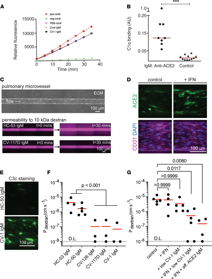
Figure 5. Features of anti-ACE2 IgM antibodies.
(A) Anti-ACE2 IgM antibodies from patient CV-1 or control do not inhibit ACE2 activity. Positive and negative controls were ACE2 alone, and ACE2 plus ACE2 inhibitor, respectively (see Supplemental Figure 6A). (B) IgM antibodies to ACE2 activate complement. Purified IgMs from anti-ACE2 IgM–positive COVID-19 patients (n = 8) and healthy controls (n = 11) was used for C1q binding assays. Values are means from 2 independent experiments performed on different days. ***P < 0.0001, Mann-Whitney test. (C–G) Anti-ACE2 IgM affects the pulmonary endothelium. (C) Phase image of a pulmonary microvessel (top). Fluorescence images of microvessels exposed to anti-ACE2–negative IgM (HC) or anti-ACE2–positive IgM (CV) after perfusion with 10 kDa dextran (lower). Representative images across n = 3 to 6 independent experiments for each IgM condition are shown. (D) ACE2 and CD31 microvessel staining following 24-hour perfusion with IFN-α/γ. Representative images across n = 3 independent experiments are shown. (E) C3c staining after perfusion with IFN and anti-ACE2–positive or control IgM (3.33 μg/mL). Representative images across n = 3 independent experiments are shown. (F) Permeability of microvessels perfused with IFN and anti-ACE2–positive IgM (CV) or anti-ACE2 negative IgM (HC) (100 μg/mL). D.L., detection limit. A linear mixed effects model was used to test the effect of anti-ACE2–positive IgM on permeability. n = 3 to 5 for each IgM condition; each dot represents an independent replicate. (G) Inhibition of microvessel permeability to 10 kDa dextran in response to anti-ACE2 IgM is IFN dependent. Low CV-1: 3.33 μg/mL; affinity-purified anti-ACE2 IgM: 100 ng/mL. To compare each condition to control, a Kruskal-Wallis test followed by Dunn’s multiple-comparison test was performed. n = 4 to 6 for each IgM condition; each dot represents an independent replicate.

