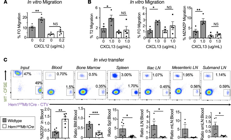
Figure 4. Conditional deletion of Hem-1 in B cells disrupts B cell migration and homing to lymphoid tissues.
In vitro migration of splenic B cells isolated from WT and Hem1fl/flMb1Cre mice in transwell plates following stimulation with 0.2 μg/mL CXCL12 or 1 μg/mL CXCL13. (A) Shown are bar graphs depicting the percent FO B cells migrated. Data points are representative of individual mice from a single experiment. Data are representative of 2 individual experiments (n = 8 and n = 8). (B) Shown are bar graphs depicting the percent T2, FO, and MZ/MZP cells migrated in the absence or presence of CXCL13 stimulation. Data points are representative of individual mice from a single experiment. Data are representative of 2 individual experiments (n = 7 and n = 7). (C) WT CFSE-labeled B cells were mixed 1:1 with CTV-labeled Hem1fl/flMb1Cre B cells combined at a 1:1 ratio and injected into WT host mice. The representation of WT and Hem1fl/flMb1Cre B cells in the input (far left) and recipient tissues (right) were determined by flow cytometry 24 hours after i.v. injection. Shown are representative dot plot histograms (top) and graphical representations (bottom) of the ratios of B220+ B cells in peripheral blood versus each respective tissue in individual recipient mice. The data are representative of 2 independent experiments of recipient mice (n = 10). Data represent the mean ± SEM and were analyzed via an paired Student’s t test (A and B) and unpaired Students t test (C). *P < 0.05, **P < 0.01, ***P < 0.001.

