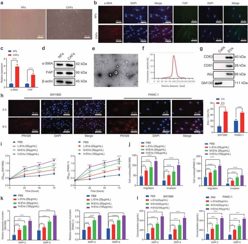
Figure 1.

CAFs-derived EVs promote the proliferation, migration and invasion of PC cells.
A. Inverted microscopic images of the morphologies of CAFs and NFs. B. IF detection of the expression of α-SMA and FAP in CAFs and NFs. C. Expression of α-SMA and FAP mRNAs in CAFs and NFs determined by RT-qPCR. D. Expression of α-SMA and FAP proteins in CAFs and NFs determined by Western blot analysis. E. TEM visualization of the morphology of CAFs-derived EVs. F. Size distribution of the CAFs-derived EVs assessed by NTA. G. Expression of EVs-related proteins determined by Western blot analysis. H. Internalization of EVs by SW1990 and PANC-1 cells under a confocal microscope. Blue indicates DAPI staining and rad indicates PKH67-labeled EVs. I. The proliferation of SW1990 and PANC-1 cells after co-culture with CAFs-derived EVs for 48 h measured by CCK-8 assay. J. Migration and invasion of SW1990 and PANC-1 cells after co-culture with CAFs-derived EVs for 48 h measured by Transwell assay. K. Expression of tumor-related proteins MMP-2 and MMP-9 in SW1990 and PANC-1 cells after co-culture with CAFs-derived EVs for 48 h determined by Western blot analysis. L. Expression of MMP-2 and MMP-9 in the supernatant of SW1990 and PANC-1 cells following co-culture with EVs for 48 h measured by ELISA. Experiments were repeated 3 times. ** indicates p < .01, *** indicates p < .001, and **** indicates p < .0001.
