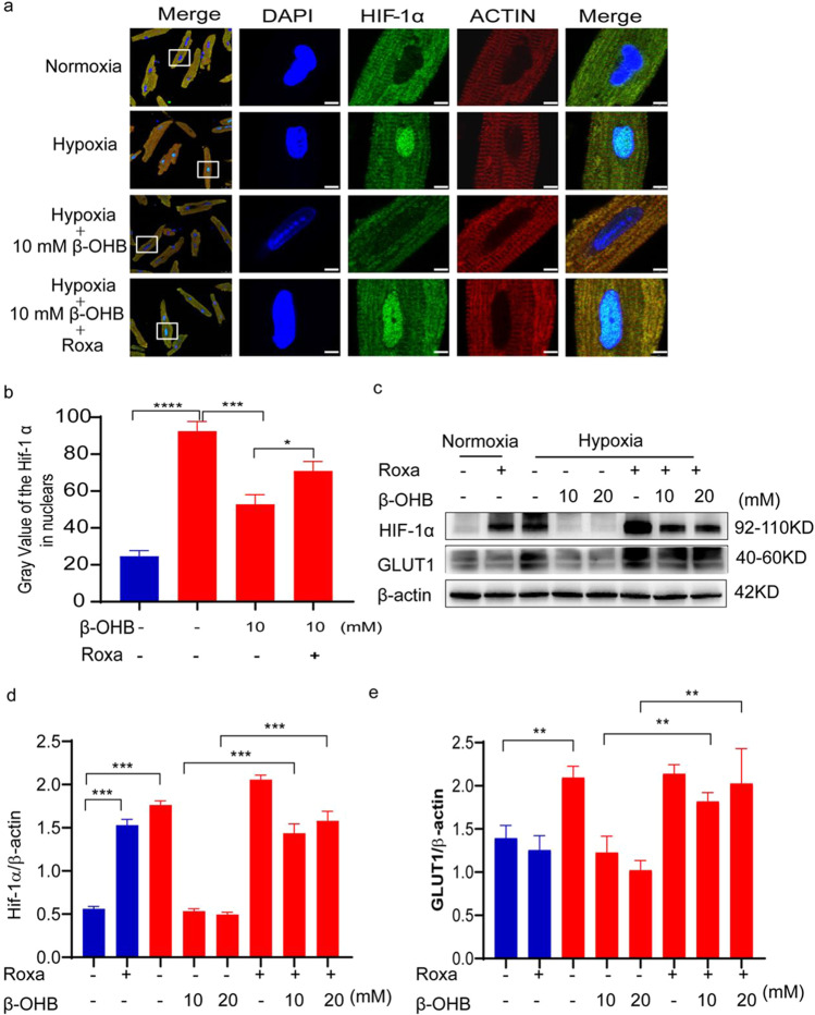
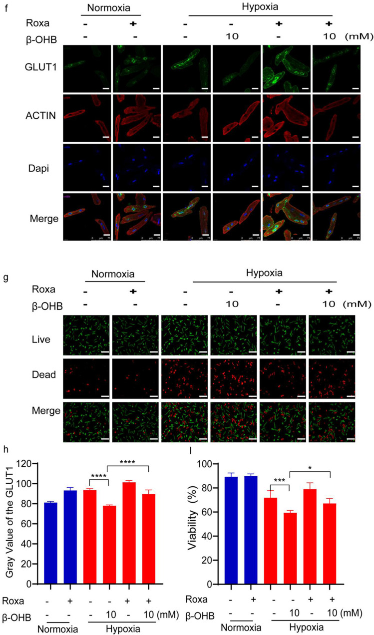
Fig. 6.
Roxadustat partially reversed the effects of β-OHB in cardiomyocytes under hypoxic conditions. a Immunofluorescence imaging showing HIF-1α expression in cardiomyocytes cultured with 50 μM roxadustat and 10 mM β-OHB under normoxia or hypoxia for 12 h, scale bar, 5 μm; b quantitation of HIF-1α in the nuclei of CMs cultured with 50 μM roxadustat and 10 mM β-OHB under normoxia or hypoxia for 12 h; c, d, and e western blot of HIF-1α and GLUT1 expression in cardiomyocytes cultured with 50 μM roxadustat and β-OHB (10, 20 mM) under normoxia or hypoxia for 12 h (n = 3 in each group); f immunofluorescence imaging of GLUT1 in CMs cultured with 50 μM roxadustat and 10 mM β-OHB under normoxia or hypoxia. Scale bars, 25 μm; g live (green) or dead (red) CMs cultured with 50 μM roxadustat and 10 mM β-OHB under normoxia or hypoxia, scale bars, 50 μm; h quantitation of GLUT1 in CMs cultured with roxadustat and 10 mM β-OHB under normoxia or hypoxia for 12 h (n = 3 in each group); i quantitation of viability of CMs cultured with roxadustat and 10 mM β-OHB under normoxia or hypoxia. Data are mean ± SEM. *P < 0.05, **P < 0.01, ***P < 0.001, ****P < 0.0001


