Figure 3.
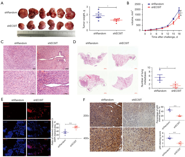
Knockdown of the expression of ECSIT in MDA-MB-231 cells suppressed tumorigenesis and metastasis and increased cell death in tumor bearing mice. Control and ECSIT knockdown stable cell lines were injected subcutaneously into nude mice. The tumor sizes were measured every three days until they reached 2 cm3. (A) Tumor size and tumor weight are shown (n=6). (B) Tumor growth curves of MDA-MB-231 cells in female nude mice are presented (n=6). (C) Representative H&E slides from breast cancer in nude mice 18 days after subcutaneous injection of breast cancer cells. (D) Circles indicate metastatic lesions in the left panels. Quantification of microscopic MDA-MB-231 lung metastases 18days after subcutaneous tumor cell injection (right panel). (E) Immunofluorescence staining for cleaved caspase-3 was performed to analyze cell death in xenograft tumors (left panel; scale bar =50 µm). Quantification of cleaved caspase-3 positive (right panel; n=6) within tumor slides and corresponding representative images of each staining. (F) Representative IHC staining of tumors harvested 18 days after injection. The brown granulation shows p65 expression and nuclei counterstained with hematoxylin (blue). Scale bars on the top indicate 50 µm and scale bars below indicate 20 µm. Quantification of p65 positive (n=6) within tumor slides and p65 staining scores were measured by ImageJ with an IHC Profiler. Statistical significance was calculated for the indicated paired samples with *, P<0.05 and ***, P<0.001. shRandom, negative control; shECSIT, knockdown of ECSIT.

